Sonoscape Ultrasound
- Product Introduction
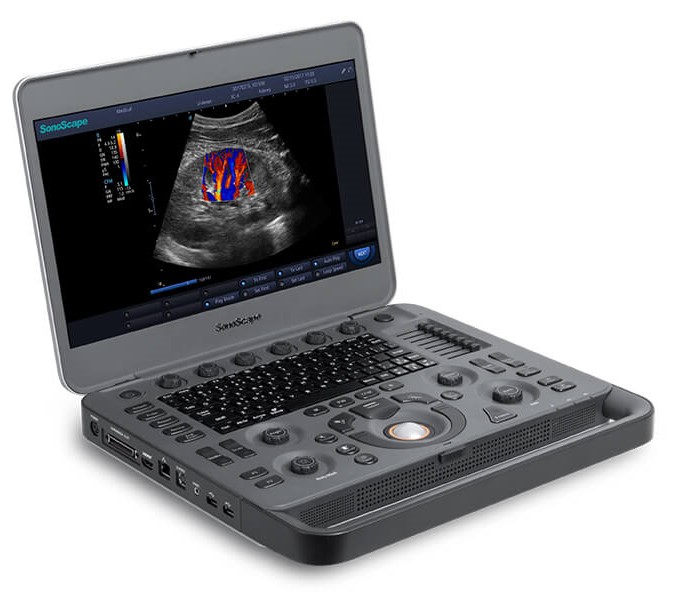
Hardware:
X3 Main Unit
15.6" High Resolution LED Color Monitor (with Auto-adaptive LED Backlight)
One Transducer Port
USB 2.0 / Hard Disk 500 G
Standard Battery
WiFi Module
Adaptor
Software:
B ( 2B & 4B ) Mode
M Mode
Color Doppler Flow Imaging
Power Doppler Imaging / Directional Power Doppler Imaging
Pulse Wave Doppler Imaging
Continuous Wave Doppler Imaging
Dynamic Multi-beam Technology
Tissue Harmonic Imaging
Pure Inversion Harmonic Imaging
Tissue Specific Imaging
Compound Imaging
Widescan: Linear Extended
Widescan: Convex Extended
μ-Scan (2D Speckle Reduction Technology)
Auto Trace
TEI Index
One-Key Optimization (Auto Optimization for 2D / M / PW / CW)
TGC (Time Gain Compensation)
LGC (Lateral Gain Compensation)
SR Flow
Simultaneous Mode (Triplex)
B / C Dual Live
Standby Mode
B Mode Panoramic Imaging
Show Gallery
2D Steer
Vis-Needle (Needle Visualization Enhancement)
Auto IMT
ECG (Software: Must be Configured with I/O Docking Extender )
DICOM 3.0: Store / C-Store / Worklist / MPPS / Print / SR / Q&R
X3 Optional Configurations:
CFM M Mode
Tissue Doppler Imaging
Tissue Doppler Imaging with M Mode
Anatomic M Mode
Prospective Saving
Probe Choice:
Linear Array L741
Linear Array 9L-A
Convex Array 3C-A
Convex Array C322
Micro-convex Array C613
Phased Array 3P-A
Phased Array 7P-B
Endocavity 6V1
Endocavity EC9-5
Linear Array 10I2
Convex Array 6CT-A
CWD 2.0